 EN
ENIn modern surgery involving the arms and legs, general anesthesia is increasingly becoming optional rather than mandatory. The German Society for Ultrasound in Medicine (DEGUM) highlighted that targeted, temporary nerve blocks guided by high-resolution ultrasound allow complex surgical procedures to be performed safely while keeping patients conscious or lightly sedated. Dr. med. Karsten Pracht, Chief Physician of Anesthesiology, Intensive Care, Pain Therapy, and Palliative Medicine at Sana Kliniken Leipziger Land and President of DEGUM, explained ahead of the 49th Three-Country Meeting in Mainz that visual needle tracking fundamentally transforms regional anesthesia. This technological advancement significantly reduces cardiovascular stress, making it an invaluable option for elderly patients and individuals with pre-existing medical conditions who travel to Germany for specialized care.
Principles of Regional Anesthesia and Anatomical Variations
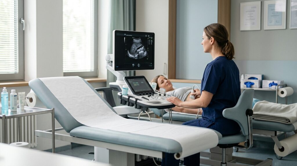
Surgical interventions on the upper and lower extremities, such as fixing bone fractures, replacing knee joints, or repairing complex ankle injuries, historically required systemic general anesthesia. General anesthesia places considerable strain on the entire organism, particularly affecting the cardiovascular system and respiratory function. Regional anesthesia was developed to block pain signals specifically at the nerve site leading from the limb to the central nervous system. By interrupting signal transmission along targeted nerve bundles, surgeons can operate effectively without subjecting the entire body to systemic narcotic agents.
For many years, anesthesiologists performed regional nerve blocks relying primarily on anatomical landmarks, surface palpation, and indirect sensations reported by the patient or felt through tactile resistance. While experienced specialists achieved reliable results, human anatomy varies significantly between individual patients. Nerve pathways do not always follow textbook paths, and adjacent structures such as blood vessels or muscle fascia can shift their relative positions based on age, body composition, or prior surgeries. These natural anatomical variations historically made precise local anesthetic placement challenging and occasionally required higher medication doses to ensure complete blockade.
The integration of real-time ultrasound technology addresses these anatomical variations directly by rendering internal structures visible before any puncture takes place. Before inserting a needle, the anesthesiologist can scan the exact area, map out the trajectory, and identify unique anatomical features specific to that individual patient. This visual mapping ensures that nerve bundles are accurately located regardless of individual variations or previous structural changes caused by disease or injury.
For international patients considering surgical treatment in Germany, understanding these foundational principles highlights why diagnostic and procedural visualization is so crucial. Modern anesthesia focuses not only on eliminating pain during surgery but also on minimizing systemic toxicity and physiological stress. By replacing blind or landmark-based techniques with direct visual guidance, medical centers can offer localized pain control tailored precisely to the patient’s unique anatomy.
High-Precision Guidance and Reduced Medication Doses
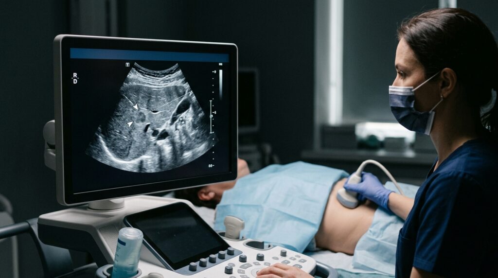
During ultrasound-guided regional anesthesia, the anesthesiologist observes the continuous movement of the puncture needle live on a high-resolution screen throughout the entire procedure. As Dr. med. Karsten Pracht emphasized, seeing the exact course of a nerve and the surrounding structures allows specialists to guide the needle with millimeter precision. This live visual feedback prevents accidental needle contact with adjacent blood vessels, sensitive soft tissues, or the nerve trunk itself. Avoiding intravascular injection of local anesthetics is a critical safety factor that drastically reduces the risk of systemic toxic reactions.
Another major advantage of real-time sonographic monitoring is the ability to observe the actual distribution of the anesthetic fluid as it is injected around the nerve sheath. In traditional landmark techniques, larger volumes of local anesthetic were frequently administered to compensate for slight positioning uncertainties and ensure the drug spread sufficiently to block pain signals. With direct ultrasound visualization, physicians can confirm that the liquid envelopes the target nerve completely using much smaller medication volumes.
According to DEGUM President Dr. med. Karsten Pracht, working with higher accuracy and reduced drug dosages substantially improves both the reliability and overall safety of regional anesthesia concepts. Lower doses mean reduced metabolic burden on the liver and kidneys, which process and clear these medications from the bloodstream. Furthermore, targeted delivery ensures that neighboring nerves responsible for uninjured muscle groups remain unaffected whenever possible, preserving motor control in non-target areas.
The technological advancements outlined by DEGUM illustrate why German clinical centers maintain high standards in perioperative sonography. The 49th Three-Country Meeting in Mainz showcases these standards, highlighting specialized training sessions on ultrasound-guided vascular access and neuroaxial procedures. For patients undergoing procedures in Germany, this translates into standardized clinical protocols executed by highly trained anesthesiologists who utilize state-of-the-art ultrasound imaging in daily practice.
Advantages for High-Risk Patients and Postoperative Recovery

The benefits of ultrasound-guided nerve blocks are particularly pronounced in older patients and those with serious underlying chronic conditions. General anesthesia carries heightened risks for individuals with heart disease, chronic obstructive pulmonary disease, renal insufficiency, or neurological vulnerabilities. Localized nerve blockade maintains hemodynamic stability without depressing cardiac output or respiration. Consequently, elderly patients experience far less postoperative confusion, cognitive decline, or cardiovascular strain, facilitating a much smoother and safer recovery process.
Beyond serving as a complete substitute for general anesthesia, regional ultrasound techniques are frequently combined with general anesthesia for major complex surgeries. When a targeted nerve block is applied alongside general anesthesia, the patient requires significantly lower doses of systemic anesthetic agents and intravenous painkillers during the operation. Postoperatively, this combined approach dramatically reduces the need for heavy opioid analgesics, thereby minimizing common opioid-related side effects such as severe nausea, vomiting, bowel dysfunction, and sedation.
Effective, targeted pain relief directly supports modern fast-track rehabilitation protocols used in German hospitals. By controlling surgical pain without inducing total motor paralysis or systemic lethargy, patients can begin early mobilization shortly after joint replacements or fracture repairs. Early movement reduces the risk of deep vein thrombosis, pulmonary embolism, and muscle atrophy, enabling shorter hospital stays and faster return to independence, which is especially valuable for international patients who wish to travel safely back home.
Patients preparing for orthopedic or extremity surgery in Germany are encouraged to discuss regional anesthesia options with their attending medical team during preoperative consultations. Inquiries regarding whether ultrasound guidance can be utilized for their specific intervention, how pain management will be structured after surgery, and whether a combined approach is recommended can help personalize the treatment plan. Understanding these options empowers patients and their families to make informed decisions alongside German medical specialists.
Source: Deutsche Gesellschaft für Ultraschall in der Medizin (DEGUM)