 EN
Surgeons at Universitätsklinikum Leipzig have integrated artificial intelligence, three-dimensional digital modeling, and augmented reality into robotic surgery for kidney tumors. By projecting a personalized 3D virtual hologram directly into the display of the Da-Vinci surgical robot, physicians can visually navigate through delicate renal tissue in real time. A multi-center trial led by Universitätsklinikum Leipzig across ten major medical centers in Germany confirmed that this innovative navigation method significantly enhances surgical precision, reduces procedure time, and doubles the rate of tissue-sparing vascular clamping. For patients suffering from localized kidney neoplasms, this technological step forward offers improved preservation of healthy renal tissue without compromising oncological safety.
Kidney Tumors and Organ-Preserving Surgery in Germany
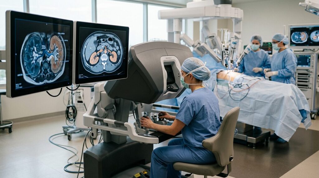
Malignant neoplasms of the kidney represent a serious oncological challenge, yet early diagnosis frequently allows for favorable therapeutic outcomes. When a kidney tumor is discovered at a localized stage before regional or distant metastasis has occurred, radical or partial surgical removal yields five-year survival rates between 80 and 90 percent. However, complete removal of an entire kidney can lead to a long-term decline in overall renal function, increasing the future risk of chronic renal failure and cardiovascular complications. Consequently, modern urological surgery prioritizes nephron-sparing approaches whenever technically feasible.
Diagnostic evaluation of suspected renal masses relies primarily on contrast-enhanced computed tomography (CT) and magnetic resonance imaging (MRI). These radiological modalities provide detailed two-dimensional cross-sectional anatomical images of the tumor, its precise anatomical dimensions, and its proximity to major blood vessels and the urinary collecting system. Physicians analyze these static scans prior to entering the operating room to formulate a comprehensive surgical plan tailored to the specific anatomical features of each patient’s kidney.
To execute complex organ-preserving operations with high precision, specialized urological centers in Germany frequently utilize minimally invasive robotic platforms such as the Da-Vinci surgical system. The operating surgeon controls articulated instruments from a dedicated console while viewing a magnified three-dimensional image of the operative field. During traditional robotic procedures, surgeons may also apply intraoperative ultrasound by placing a small flexible probe directly onto the surface of the kidney. This ultrasound probe provides real-time cross-sectional images, assisting the surgical team in locating deep-seated lesions beneath the organ’s surface.
Augmented Reality and the German Multi-Center Study
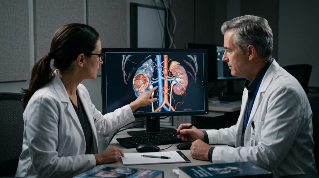
The new surgical approach developed and evaluated at Universitätsklinikum Leipzig combines high-resolution imaging with artificial intelligence to generate dynamic three-dimensional anatomical models. Patient CT and MRI data are processed by specialized software that creates a digital reconstruction of the affected kidney. In this interactive model, neoplastic tissue, arterial and venous vascular trees, and healthy parenchyma are color-coded and spatially delineated. Operating surgeons can rotate the virtual model on screen prior to surgery, hide or isolate specific anatomical structures, and chart the safest surgical route to the tumor before making the initial incision.
The key technological breakthrough occurs during the surgical procedure through the application of augmented reality. Inside the display console of the Da-Vinci robot, the preoperatively generated 3D model is projected as a virtual hologram overlaying the actual surgical view. This hologram aligns with millimeter precision over the physical organ, effectively allowing the surgeon to see through the surrounding tissue. As a result, concealed blood vessels and exact tumor margins become visible in real time, granting surgeons superior spatial orientation before cutting into the renal tissue.
The tangible clinical benefits of this technology were demonstrated in a nationwide clinical study funded by Deutsche Krebshilfe. Conducted between October 2023 and October 2025 under the leadership of the urology department at Universitätsklinikum Leipzig, the trial evaluated 370 patients across ten major German hospitals. This study represents the largest clinical investigation worldwide comparing conventional two-dimensional imaging against novel three-dimensional augmented reality navigation during robotic kidney surgery.
The study results demonstrated significant clinical advantages for patients managed with 3D augmented reality visualization. Surgeons using the technology spent significantly less time operating at the robotic console compared to traditional procedures. Furthermore, the rate of selective clamping—a delicate technique where only the specific blood vessels directly supplying the tumor are occluded while blood flow and oxygenation continue to the rest of the kidney—increased from 9.2 percent in the control group to 19.4 percent in the 3D group. Surgical feedback was overwhelmingly positive, with 93 percent of participating surgeons expressing satisfaction or high satisfaction with the quality and utility of 3D image guidance.
What Advanced 3D Surgery Means for Patients

Preserving healthy renal tissue is of vital importance for long-term health, renal filtration capacity, and general quality of life. Prof. Jens-Uwe Stolzenburg, Director of the Klinik und Poliklinik für Urologie am Universitätsklinikum Leipzig, emphasizes that combining robotic surgery, 3D digital modeling, and real-time augmented reality represents a major milestone toward navigated minimally invasive urology. By enabling precise identification of tumor margins, the technology allows surgeons to maximize functional kidney preservation while ensuring complete oncological resection.
Universitätsklinikum Leipzig has long maintained a pioneering role in the adoption of state-of-the-art surgical technologies in Germany. Prof. Jens-Uwe Stolzenburg was the first surgeon at UKL to introduce the Da-Vinci robotic system, establishing a center of excellence for minimally invasive organ-sparing renal surgery. The integration of artificial intelligence and real-time hologram navigation continues this commitment to clinical innovation, setting new standards for urological care across German university hospitals.
International patients seeking specialized treatment in Germany for diagnosed kidney tumors can benefit directly from these technological innovations. The 3D augmented reality system is already fully implemented in clinical routine at Universitätsklinikum Leipzig and is being progressively adopted by leading urological centers. When consulting with urological specialists in Germany, patients and their families are encouraged to inquire whether 3D modeling, augmented reality visualization, and selective vascular clamping are suitable for their specific anatomical condition.
Source: Universitätsklinikum Leipzig AöR