 EN
A comprehensive review published in Deutsches Ärzteblatt International and featured at the 31st Annual Meeting of the Deutsche Assoziation für Fuß und Sprunggelenk (D.A.F.) in Bonn marks a major shift in how ankle fractures are evaluated and treated. Experts from German university hospitals and specialized orthopedic institutions, working under the umbrella of the Deutsche Gesellschaft für Orthopädie und Unfallchirurgie (DGOU), emphasize that a broken ankle no longer automatically implies surgical intervention or six weeks of mandatory rest. Current clinical evidence demonstrates that mechanically stable fractures can be managed safely without an operation, while early pain-adapted weight-bearing after necessary surgery accelerates functional recovery without raising complication rates.
Understanding Ankle Fractures and Modern Diagnostic Precision
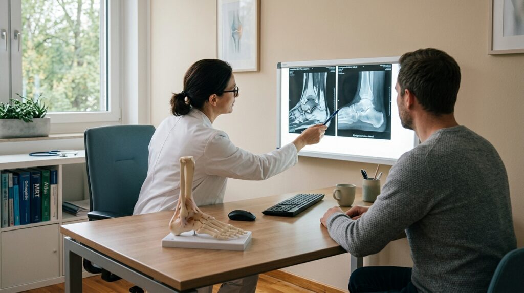
Ankle fractures represent one of the most common acute skeletal injuries in adults, with an incidence of approximately 74 cases per 100,000 inhabitants every year. The ankle joint connects the lower leg to the foot and plays a fundamental role in maintaining stability and mobility during walking. A fracture can involve the tibia, the fibula, or both bones simultaneously, and is frequently accompanied by damage to stabilizing ligament structures such as the deltoid ligament or the syndesmosis. Everyday accidents, such as twisting an ankle during athletic activities, misstepping on stairs, or slipping on uneven ground, are common causes of these painful injuries.
Restoring precise anatomical alignment of the joint is critical for long-term health, as even minor joint incongruity or residual instability can impair mobility over time. Uneven weight distribution across damaged articular surfaces accelerates mechanical wear, significantly increasing the risk of secondary post-traumatic osteoarthritis. As primary study author Prof. Dr. Sebastian Felix Baumbach notes, although ankle fractures are often regarded as routine clinical events, long-term follow-up data show that one in five patients continues to suffer from substantial complaints years after the injury. For this reason, German specialists prioritize meticulous initial diagnostic evaluations to identify subtle joint instabilities early.
Diagnostic protocols in German specialized centers have advanced far beyond traditional static two-dimensional X-rays to assess whether a fracture is truly stable. Physicians routinely utilize stress radiographs under functional load together with high-resolution computed tomography to evaluate the mechanical integrity of the syndesmotic complex and surrounding structures. This detailed imaging strategy enables orthopedists to differentiate stable injuries from complex instable patterns with high precision. By confirming structural stability under functional conditions, clinicians can avoid unnecessary surgical operations and design tailored conservative rehabilitation programs tailored to each patient’s needs.
Re-Evaluating Surgical Indications and Early Weight-Bearing
Traditional treatment algorithms historically mandated surgical fixation for a broad range of ankle fractures, followed by six weeks of absolute non-weight-bearing in a rigid cast. However, the scientific review published in Deutsches Ärzteblatt International by lead author Prof. Dr. Sebastian Felix Baumbach and senior author Prof. Dr. Stefan Rammelt, titled "Diagnose, Behandlung und Nachbehandlung von Sprunggelenkfrakturen" ("The diagnosis, treatment, and aftercare of ankle fractures"), challenges this rigid historical approach. Their research establishes that when dynamic joint stability is verified through modern imaging, conservative non-surgical management achieves excellent clinical outcomes while sparing patients from operative risks.
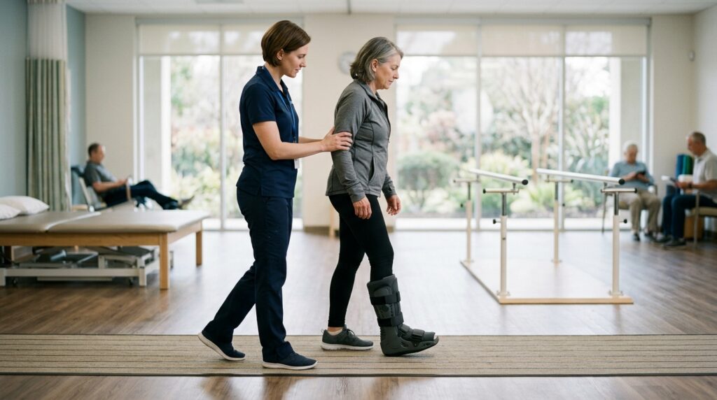
For patients who do require surgical realigning and internal fixation, post-operative rehabilitation protocols are experiencing an equally significant evolution. In a dedicated multicenter study titled "Schmerzadaptierte Vollbelastung nach operativ versorgten Sprunggelenksfrakturen" ("Pain-Adapted Full Weight-bearing After Ankle Fracture Surgery"), conducted across three university hospitals in Germany, researchers investigated early mobilization protocols. The investigative team, including Prof. Dr. Sebastian Felix Baumbach, Prof. Dr. T. Mittlmeier, Dr. D. C. Fischer, Prof. Dr. W. Böcker, Prof. Dr. Stefan Rammelt, and Prof. Dr. H. Polzer, proved that early pain-adapted full weight-bearing after surgery is clinically safe and does not increase hardware or wound complications.
Highlighting the clinical importance of these findings, lead author Prof. Dr. Sebastian Felix Baumbach stated: "Die frühzeitige, schmerzadaptierte Vollbelastung ist sicher und stellt eine deutliche Verbesserung der Lebensqualität der Betroffenen dar" ("Early, pain-adapted full weight-bearing is safe and represents a significant improvement in the quality of life for those affected"). Allowing patients to load the injured leg according to their personal pain tolerance reduces joint stiffness, preserves calf muscle mass, and speeds up the return to normal daily routines. Consequently, the rigid historical standard of six weeks of mandatory unloading is being replaced by flexible, patient-centered rehabilitation models.
Developing S3 Guidelines and Practical Patient Guidance

To translate these scientific advances into standardized clinical practice across hospitals, an interdisciplinary panel of medical experts is currently developing a new S3 guideline titled "Evidenzbasierte Diagnostik und Therapie von Sprunggelenkfrakturen (EviDenTS)". Supervised by the DGOU and D.A.F., this authoritative guideline is scheduled for official publication by December 31, 2027. Dr. Jörn Dohle, Vice President of DGOU, emphasized that treatment strategies have progressed markedly in recent years, making formal evidence-based guidelines essential to ensure that every patient receives optimized, modern care grounded in high-level research.
The practical implementation of these diagnostic and therapeutic insights was a central topic at the 31st Annual Meeting of D.A.F., held from September 10 to 12, 2026, in Bonn. Leading foot and ankle surgeons, traumatologists, and rehabilitation specialists gathered to refine treatment pathways that combine advanced imaging with early controlled weight-bearing. By establishing unified, evidence-based clinical recommendations, German orthopedic institutions aim to reduce the incidence of long-term joint pain and improve overall long-term functional recovery for domestic and international patients alike.
International patients seeking orthopedic care or surgical consultation in Germany can benefit directly from these updated, evidence-driven management concepts. Although the formal EviDenTS S3 guideline will be completed by late 2027, specialized German hospitals already integrate these principles into daily clinical care. Patients undergoing evaluation should discuss with their attending German specialist whether their fracture pattern is mechanically stable, whether computed tomography or stress imaging is recommended, and how an early pain-adapted weight-bearing plan can be structured safely for their specific injury.
Source: Deutsche Gesellschaft für Orthopädie und Unfallchirurgie e. V.