 EN
ENThe United States Food and Drug Administration (FDA) has granted official approval to 18F-Fluorethyltyrosin, known as FET, a diagnostic radiotracer developed at Forschungszentrum Jülich in Germany. Marketed under the name Pixclara® by Telix Pharmaceuticals, this specialized radioactive substance marks a major milestone in neuro-oncology imaging, particularly for evaluating patients previously treated for gliomas. For international patients seeking advanced care in Germany, this milestone highlights a sophisticated diagnostic tool that helps physicians distinguish between recurrent tumor growth and benign post-treatment changes. Understanding whether brain tissue alterations stem from active cancer or treatment effects is critical for choosing the right next therapeutic steps.
Gliomas and the Diagnostic Challenges After Brain Tumor Therapy

Gliomas are among the most common primary brain tumors, originating from the supportive glial cells of the central nervous system. Treatment for high-grade or complex gliomas typically involves a combination of surgical resection, radiotherapy, and systemic therapies such as chemotherapy. While these medical interventions are designed to control tumor growth, they also induce significant structural and chemical changes within the surrounding healthy brain tissue. As a result, monitoring a patient after primary treatment requires highly sensitive diagnostic methods to ensure that any suspicious physical changes in the brain are accurately evaluated.
In routine neuro-oncological practice, magnetic resonance imaging (MRI) serves as the primary tool for detecting brain tissue changes. MRI provides detailed anatomical pictures of brain structures, enabling physicians to observe physical alterations over time. However, conventional MRI has a well-known diagnostic limitation following surgery or radiation therapy. Radiotherapy and chemotherapy often cause localized tissue inflammation, scar formation, or radiation necrosis, which frequently appear virtually identical to an actively growing brain tumor on standard structural scans.
This diagnostic ambiguity creates a clinical dilemma for patients and multi-disciplinary medical teams. If a doctor mistakes treatment-related tissue changes for a recurring tumor, the patient might undergo unnecessary secondary surgery or aggressive toxic treatments. Conversely, mistaking a true tumor recurrence for harmless treatment effects can delay essential, lifesaving therapeutic adjustments. Resolving this diagnostic uncertainty requires specialized functional imaging techniques that look beyond simple anatomical structure and instead evaluate the biological activity and metabolic behavior of brain tissue.
The Science Behind FET and its Decades of Research at Jülich
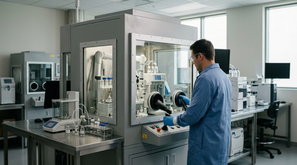
To address this diagnostic challenge, researchers at Forschungszentrum Jülich developed 18F-Fluorethyltyrosin, abbreviated as FET. FET is a synthetic radiotracer designed to mimic a natural amino acid, chemically labeled with a minute quantity of radioactive fluorine-18. Because malignant glioma cells consume amino acids at a significantly higher rate than healthy brain cells, FET accumulates selectively within active tumor tissue. When a patient receives FET, a Positron Emission Tomography (PET) scanner captures the precise locations of radiotracer accumulation, providing physicians with metabolic mapping of suspicious brain lesions.
The development and clinical validation of FET-PET reflect decades of rigorous scientific work at Forschungszentrum Jülich. Research on FET began in the 1990s and intensified around the turn of the millennium under the leadership of Prof. em. Dr. Karl-Josef Langen. Multiple specialized units collaborated on the project, including the Institute of Neuroscience and Medicine’s Cognitive Neuroscience (INM-3), Physics of Medical Imaging (INM-4), and Nuclear Chemistry (INM-5). Working closely with university medical centers, the team published more than 170 scientific studies and evaluated more than 11,000 patients with brain tumors.
PD Dr. Philipp Lohmann, interim director of INM-4 and successor to Prof. Langen as head of the Digital Translational Neuroimaging working group, emphasized that the FDA approval of Pixclara® underscores the global impact of this long-standing interdisciplinary research. By granting approval for Telix Pharmaceuticals’ commercial formulation, the US regulator confirms the clinical value of FET-PET for evaluating complex post-treatment glioma findings. This recognition confirms that analyzing tissue metabolism alongside structural MRI yields far greater diagnostic precision than relying on anatomical imaging alone.
What This Means for Patients Seeking Specialized Treatment in Germany

It is important for patients and their families to understand that FET is strictly a diagnostic radiotracer, not a therapeutic drug. FET does not shrink tumors or directly treat cancerous tissue; rather, it provides vital functional information about tumor metabolism to guide treatment planning. In Germany, FET-PET is already integrated into specialized clinical practice, particularly through the Joint Federal Committee (Gemeinsamer Bundesausschuss, G-BA) guidelines for specialized outpatient care (ambulante spezialfachärztliche Versorgung). This framework covers complex cases where post-treatment MRI findings remain inconclusive.
At Forschungszentrum Jülich, clinical FET-PET examinations are performed at INM-4, which functions as an outpatient branch of the Department of Nuclear Medicine at Uniklinik RWTH Aachen. Medical specialists within a radius of more than 300 kilometers regularly consult the expertise of the Institute of Neuroscience and Medicine for challenging neuro-oncological cases. Although the regulatory mechanism in Germany differs from the formal drug approval pathway chosen by the FDA in the United States, German centers offer vast clinical experience and specialized infrastructure for performing and interpreting these complex scans.
For international patients traveling to Germany for neuro-oncology consultations, FET-PET represents a highly refined diagnostic option when standard MRI scans leave lingering questions. Ongoing research at Forschungszentrum Jülich continues to advance the field by combining FET-PET with hybrid imaging modalities and artificial intelligence to enhance diagnostic accuracy further. Patients discussing their care with German specialists should ask whether advanced metabolic imaging such as FET-PET is appropriate for their specific case, particularly if previous scans show ambiguous structural changes after surgery or radiation.
Source: Forschungszentrum Jülich