EN
ENOptic nerve atrophy treatment in Germany targets the cause: the atrophy is neither a verdict nor a disease in itself but the end result of damage. The fibres that carry the image to the brain have died; the doctor sees this as a pale optic disc. Patients notice not blindness but blurred sight, gaps in the visual field and faded colours.
To be clear: dead fibres cannot be restored, in Germany or anywhere else. The trip is about finding the cause, stopping ongoing damage and making the most of the remaining sight, not about «regenerating the nerve».
Why the nerve atrophies
The nerve atrophies when another disease destroys its fibres: glaucoma, whose main risk factor is raised eye pressure; inflammation of the nerve, including in multiple sclerosis; poor blood supply; a tumour or aneurysm (a bulging vessel) pressing on the visual pathway; injury; poisoning, for example by methanol or certain medicines; vitamin B12 deficiency; and inherited optic neuropathies.
The cause matters more than the atrophy because it decides the treatment: glaucoma and inflammation are treated differently, compression needs a neurosurgeon’s opinion, an inherited form means monitoring and concerns the family; see also eye hospitals in Germany.
How the cause is looked for in Germany
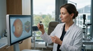
Testing goes step by step, from the eyes to, if needed, the brain and blood. Acuity testing shows how small a letter the eye can read, perimetry which parts of the visual field are missing; the nerve head is examined and eye pressure measured. Optical coherence tomography (OCT), a painless light scan, measures the nerve fibre layer, showing how many fibres remain; visual evoked potentials show how the signal reaches the brain.
Beyond the eye, an MRI of the brain and orbits shows whether a tumour or vessel is pressing on the nerve or there are inflammatory lesions as in multiple sclerosis. Blood tests look for inflammation, infection and vitamin deficiency, genetic tests for an inherited form.
A baseline matters because ongoing damage only shows in comparison: identical measurements, say, six months apart mean stability, a thinner fibre layer an active cause. Scans from home are often missing or, from other devices, not directly comparable, so the clinic usually repeats them.
What can be changed and what cannot
Dead fibres cannot be brought back, but what destroys the rest can be tackled. The retinal nerve cells that form them do not renew and barely repair themselves. Protecting or replacing them (neuroprotection, cell therapy) is being studied, but a review of this research finds both far from routine care: a paid offer to «restore the nerve» outside a clinical trial calls for caution. See what not to expect from stem cell treatment for the eye.
What can be changed is the cause: lowering the pressure in glaucoma, settling inflammation, relieving compression, correcting a deficiency, stopping a toxic drug with the prescribing doctor. The earlier, the more fibres survive.
Treatment by cause
Treatment follows the cause, and the aim is always to keep what is left. In glaucoma, eye pressure is lowered, most often with drops, otherwise by laser or surgery. Lost visual field will not fully return, but the disease can be stopped or slowed, as the DOG and BVA glaucoma guideline also sets out.
Acute optic neuritis, an inflammation of the nerve, is treated with high-dose corticosteroids, anti-inflammatory hormone drugs. Such inflammation is commonly a sign of autoimmune disease of the nervous system, above all multiple sclerosis, so tests continue and a neurologist takes over care. A tumour or aneurysm pressing on the nerve is treated with neurosurgeons without delay, as fibres keep dying while the pressure lasts; a common example is a pituitary adenoma.
In the vascular form, giant cell arteritis (inflammation of the arteries) is ruled out first; then blood pressure, sugar and lipids are controlled, with neck artery scans only when indicated. Inherited forms are confirmed genetically and monitored. For one of them, Leber hereditary optic neuropathy, the EU has authorised idebenone for adolescents and adults with visual impairment, under exceptional circumstances, as the disease is rare and full data are lacking.
Using the sight that remains
Low vision rehabilitation helps use the remaining sight: magnifiers and training with them, bright lighting and strong contrast at home and on screens, practice in using the surviving parts of the visual field, also when the centre is lost. In Germany, low vision clinics at eye hospitals provide it.
Practical questions follow: driving, study and work, disability paperwork, family support. The answers rest on measured acuity and visual field, while the rules depend on the country of residence. A retinal condition, if present, is treated in parallel, since it takes away more of the already reduced sight; see retinal treatment in Germany.
Frequently asked questions
Can optic nerve atrophy be cured?
No: no clinic or method can restore dead optic nerve fibres today. Doctors treat the cause: glaucoma pressure, inflammation, compression or a vitamin deficiency. If the cause is found and stopped in time, the remaining fibres can be preserved; how much sight remains depends on how many fibres were already lost.
Does optic nerve atrophy need surgery?
The atrophy itself is not operated on: no operation brings dead fibres back. Surgery may treat the cause, such as glaucoma that drops and laser cannot control or a tumour like a pituitary adenoma pressing on the nerve; inflammatory, vascular and inherited forms usually need none.
Is optic nerve atrophy hereditary?
Sometimes: inherited optic neuropathies such as Leber hereditary optic neuropathy are confirmed by genetic testing, and relatives may then be offered testing and counselling. Atrophy caused by glaucoma, inflammation, compression or poor blood supply is not itself inherited, although glaucoma in close relatives is a known risk factor.
How AlenMed arranges assessment and treatment
AlenMed starts with a remote review of reports, images and visual field results, which point to the likely cause, and chooses a clinic to match. One specialist centre is the Tübingen University Eye Hospital, whose neuro-ophthalmology clinic covers inflammatory, vascular, tumour-related and inherited optic nerve diseases. We send a written estimate, book the appointment, arrange the visa invitation, meet the patient and provide an interpreter at the appointment and the findings review.
Afterwards the patient keeps a report with measurements as a baseline, a written follow-up plan with intervals, advice on accompanying treatment and a contact for questions after returning home.
This material is for general information only and is not an offer. The treatment plan is decided by a doctor after an in-person examination.
News on this topic
New Retinal Chip PRIMA Restores Central Vision in Geographic Atrophy
A newly approved photovoltaic subretinal chip offers restored visual function for patients with central vision loss from geographic atrophy due to dry AMD.