 EN
EN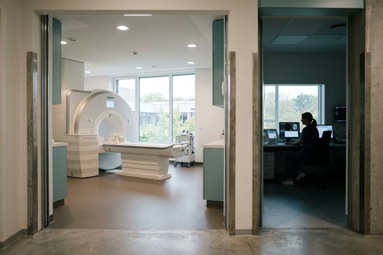
Lung cancer diagnosis in Germany begins with two questions, not with treatment: what kind of tumour this is and how far it has spread. Until both are answered, any treatment plan is guesswork; the general cancer work-up is covered on the page on cancer diagnosis in Germany.
Below, the work-up is taken step by step: what each test delivers, why steps skipped at home matter, and what can be done remotely from existing material.
Where it starts and what does not count as proof
It usually starts with a shadow on an X-ray or CT taken for another reason, or with a persistent cough, blood in the sputum, breathlessness or unexplained weight loss. Neither image nor blood test proves the diagnosis.
Tumour markers such as CEA or CYFRA 21-1 can be raised without cancer and normal with it, so the German guideline advises against them for diagnosis. Only a pathologist seeing tumour cells under the microscope confirms lung cancer; until then, “cancer” in a report is a suspicion.
A German doctor asks for the disc, not the report, and for paraffin blocks or slides, not a copy of the findings. With extra stains, “non-small cell carcinoma, not otherwise specified” may become adenocarcinoma or squamous cell carcinoma, and a lung nodule a metastasis from elsewhere. Doubtful cases involve experienced pulmonologists such as Dr Wolfgang Gesierich, medical director of Asklepios Lungenklinik Gauting.
Imaging: CT, PET-CT and the head
Imaging rests on a contrast CT of chest and abdomen: tumour size and position, lymph nodes, and any spread to vessels, pleura or chest wall. A standard CT costs approximately €500, a contrast-enhanced one around €550.
PET-CT shows metabolism, not shape: tumour cells take up labelled sugar more avidly. It is needed wherever surgery or curative radiotherapy is considered, as it finds affected nodes and distant deposits that CT misses and often changes the stage. See also PET-CT in Germany.
MRI of the head is a separate, obligatory step: the German guideline recommends it for everyone with suspected or confirmed lung cancer and treatment options, or a contrast head CT if MRI is impossible. It aims to rule out brain metastases, often silent at first yet decisive for treatment. Brain MRI with contrast costs between €800 and €1000.
How the tissue is obtained
Tissue is most often taken by bronchoscopy: a flexible tube passes through the nose or mouth after a numbing spray, with sedation if wished, and samples a tumour near the larger airways. EBUS adds an ultrasound probe for needling lymph nodes behind the airway wall from inside, without surgery, and is the first choice for central chest nodes.
A lesion at the edge of the lung is punctured through the chest wall under CT guidance and local anaesthetic. If that fails or nodes stay suspicious, surgical diagnosis follows under general anaesthetic, with a hospital stay: mediastinoscopy through a small cut above the breastbone, or thoracoscopy through small chest-wall openings. See also biopsy in Germany.
One thin core cannot carry both histology and every molecular test, so a German biopsy is planned with a margin: several samples, used sparingly, so it need not be repeated.
The molecular profile and PD-L1
The molecular profile shows which drugs may work, and it starts with one question: small cell or non-small cell cancer. They behave and are treated differently, and the pathologist tells them apart from tissue, not the radiologist from a scan.
In non-small cell cancer, gene faults with targeted drugs are sought: in advanced disease at least EGFR, ALK, ROS1, BRAF, KRAS, MET, RET, NTRK, HER2 and NRG1. PD-L1, the marker for immunotherapy prospects, is measured alongside (see immunohistochemistry in Germany). All this uses the same tissue and takes time: German guidelines allow no more than 10 working days.
Drug treatment waits for the profile, because a target can make a targeted drug the first choice instead of chemotherapy. If tissue is scarce and a repeat biopsy risky, tumour DNA is sought in the blood, a liquid biopsy. It complements tissue rather than replacing it: a negative blood result does not exclude a mutation.
Stage, tumour board and a second opinion without travelling
The stage is assembled from all results and discussed at a tumour board: in certified lung cancer centres, a thoracic surgeon, pulmonologist, oncologist, radiotherapist, radiologist and pathologist review the case together, and at LMU University Hospital in Munich, for example, the board meets every week. The report gives the stage and proposed next steps (see lung cancer treatment in Germany).
Without travelling, you can get a slide review, a second reading of images and a written remote opinion; send blocks or slides, discs and translated reports. It cannot replace an examination, lung function tests or a new biopsy.
Frequently asked questions
Can lung cancer be confirmed without a biopsy?
No: lung cancer is confirmed only by tumour tissue or cells. CT, PET-CT and tumour markers indicate probability; the tumour type shows only under the microscope. A small nodule is sometimes removed by thoracoscopy and diagnosed on that tissue: a surgical biopsy.
How long does the work-up take?
The tests usually fit into a few days if lined up in advance. The laboratory takes longer: histology first, then the molecular profile within 10 working days. Some patients stay for the board, others get the report at home; the clinic’s programme states the time frame.
Is bronchoscopy dangerous?
Serious complications are rare; the procedure itself usually takes 10 to 15 minutes. Minor bleeding is the most common problem. Sampling at the edge of the lung can let air leak between lung and chest wall, a pneumothorax that usually settles by itself or is drained.
How AlenMed arranges the work-up
Documents, slides and discs go to the clinic in advance, and AlenMed builds the programme so that tests follow one another within a few days, not weeks apart. We help with the visa invitation, and an interpreter attends appointments and the discussion of results.
Plan your stay with a margin for histology; a molecular profile that arrives later is discussed remotely. You receive images on disc and translated reports. Payment goes directly to the clinic against its cost estimate (Kostenvoranschlag); doctors’ fees follow the German GOÄ schedule, and unused advance money is refunded. A specialist consultation on existing results costs from €130, a general diagnostic package around €1,650; for treatment prices see lung cancer treatment cost in Germany.
This material is for information only and is not an offer. A doctor decides the extent of the work-up in person; final prices are set in the clinic’s cost estimate.