 EN
An arteriovenous malformation is usually found in one of two ways: after a bleed, or by chance on an MRI done for headaches or a first-ever seizure. Either way, the person hears a long word and cannot tell how urgent it is.
Arteriovenous malformation treatment in Germany starts with the scans, not with surgery. This page covers what an AVM is, why several specialists decide slowly and together, which four approaches exist and why doing nothing can also be the reasoned answer. The section page neurosurgery in Germany covers the wider field.
What an arteriovenous malformation is
An arteriovenous malformation, or AVM, is a tangle of abnormal vessels carrying blood from arteries straight into veins. Normally blood passes from an artery into capillaries, which damp the pressure, and only then into a vein. In an AVM the arteries join the veins directly through the tangle, so the veins take a pressure they are not built for.
It most often shows itself as bleeding into the brain, but it can also cause epileptic seizures or headache, and less often neurological deficits such as limb weakness or speech problems. In some people an AVM never shows itself at all.
Doctors speak of an annual bleeding risk because an AVM carries it for life. The risk depends on where the tangle, or nidus, sits, how large it is, how its draining veins are built and whether it has bled before, since a first bleed makes another more likely. No single figure fits everyone.
What is examined before the decision
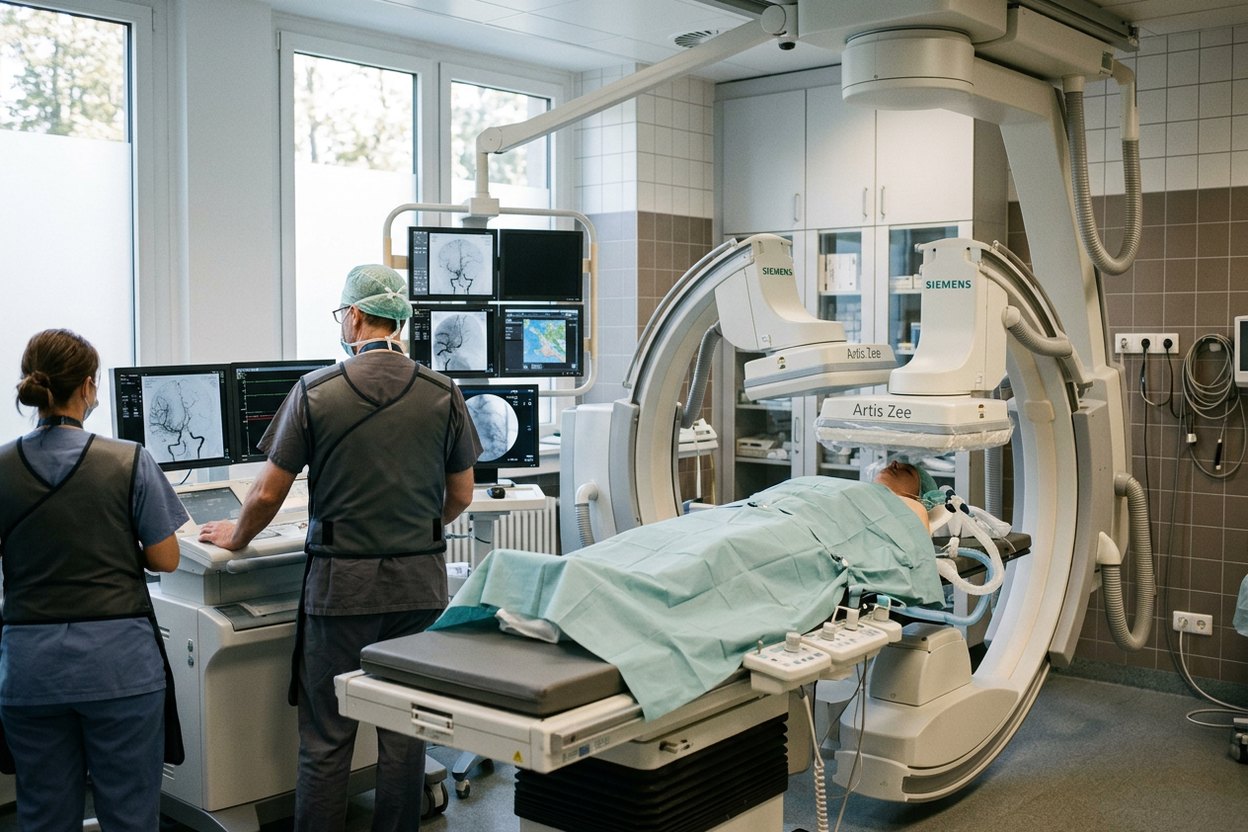
Three examinations come before the decision: MRI, CT and catheter angiography. MRI shows the brain around the malformation and traces of earlier bleeds, CT shows acute blood, and catheter angiography remains the study the plan is built on, because it shows the feeding arteries, the nidus and the drainage. The page angiography in Germany describes the procedure.
The assessment also covers where exactly the tangle lies relative to the speech and motor areas, how big it is and whether its veins drain to the surface or deep. These features are the basis of the Spetzler–Martin scale, which surgeons use to compare cases: the smaller, more superficial and further from critical areas a nidus is, the safer the operation.
In Germany such cases are discussed by a board of a neurosurgeon, a neuroradiologist and a radiation oncologist rather than by one doctor: each approach has its own specialist, and only together can they weigh the risks. It is normal for a patient to ask for the decision in writing, to reread it calmly and show it to the doctor at home.
The four approaches and how the choice is made
The choice is between observation, surgery, embolisation and radiosurgery. Observation comes first: for an unruptured malformation in a difficult location, intervention can carry more risk than the AVM itself. In the randomised ARUBA trial of unruptured AVMs, stroke or death during follow-up was less common without intervention. But as a peer-reviewed review of the ARUBA results notes, follow-up was short and many participants had embolisation alone instead of surgery, so the finding cannot simply be applied to everyone.
Microsurgical removal is the only way to take the malformation out at once and completely, and it is used where the location allows access. Embolisation closes the feeding vessels from inside through a catheter; it is more often preparation for surgery or radiosurgery, less often a treatment on its own.
Stereotactic radiosurgery is a single targeted irradiation without an incision. The vessels close not at once but over two to three years, and the bleeding risk remains throughout; this is how the gamma knife in Germany works. Methods are often combined, and there is no «best» method in general, only one that suits this nidus in this person.
After the intervention: what happens next
After surgery the patient is watched in a high-dependency bed, with blood pressure control and anti-seizure medication if there were seizures. The clinic states how long the stay lasts.
Then the result is checked: after the interval the doctor sets, a control angiography shows whether any of the nidus remains. After radiosurgery, repeated studies continue over several years until closure is confirmed.
Rehabilitation after a bleed can take months and follows the same principles as after a stroke, described on the page stroke treatment in Germany. How fully movement and speech return depends on where the bleed was and how severe it was.
Frequently asked questions
Does an arteriovenous malformation always need treatment?
No. If it has not bled and intervention in its location would be riskier than the AVM itself, doctors may advise observation: regular scans and treatment of symptoms. After a bleed, intervention is discussed more often, as another bleed is more likely. The decision rests on the scans, the symptoms and the patient’s view.
How long does arteriovenous malformation treatment take?
It depends on the approach. Surgery is one operation, then an early period in hospital and a control angiography; the clinic states the length of stay. Embolisation is sometimes staged. Radiosurgery takes one session, but the vessels close over two to three years. Rehabilitation after a bleed can take months.
How much does arteriovenous malformation treatment cost in Germany?
There is no single price: it depends on the approach and the number of stages, with examinations, intervention, hospital stay and follow-up scans charged separately. The clinic issues a written estimate once a neurosurgeon has reviewed the scans; before that, any figure would be unverified.
How AlenMed arranges AVM treatment in Germany
An enquiry starts with the scans. AlenMed sends the MRI and angiography as DICOM files, the discharge summary and a description of the seizures to a neurosurgeon; he first says whether an intervention is indicated at all and which one, and only then does the clinic issue a written estimate. Among the doctors the company works with is Prof. Erdem Güresir, director of the clinic and polyclinic for neurosurgery at Leipzig University Hospital. AlenMed works with 86 partner hospitals in 38 German cities and 345 doctors, with its office in Munich; the page neurosurgery clinics in Germany helps compare departments.
Ask in advance who exactly will operate, what is done if the nidus is not completely closed after embolisation, and who will run the follow-up studies once you are home.
This material is for information only and is not an offer. The treatment plan is decided by a doctor at an in-person consultation.