EN
Melanoma frightens people by its reputation more than by the course of the disease: an early tumour is usually cured by surgery, and advanced disease is treated with immunotherapy and targeted drugs. Most people seeking melanoma treatment in Germany arrive with a mole already removed and an alarming report that does not say what to do next. German oncology starts there, with facts about the particular tumour rather than emotions.
Below we explain how the diagnosis is rechecked, why tumour depth outweighs spot size, which methods are used and when the operation already done is enough.
What patients usually arrive with
Most patients arrive with the mole already removed and a histology report in hand. The report holds what matters in melanoma: how deep the tumour has grown into the skin and whether it was ulcerated. This, not the size of the spot by eye, decides everything that follows. For other skin tumours, such as basal cell carcinoma, see skin cancer treatment.
They travel with one question: is what has been done enough, and has anything important been missed? Was the margin wide enough, should the nodes be checked, are tests needed to choose drugs? Only the records and the tumour tissue can answer this, so doctors in Germany start by rechecking them.
How the diagnosis is rechecked in Germany
The diagnosis is rechecked in steps, starting with the slides: a German pathologist re-examines the tissue already taken, confirms that it is melanoma and re-measures the depth of invasion and the other features that set the extent of treatment. This is not bureaucracy: whether anything beyond the operation already done is needed depends on it. Slides can be sent ahead for a second opinion from a German doctor.
If the depth gives reason to check whether cells have spread, a sentinel lymph node biopsy follows: the node that lymph from the tumour reaches first is located and examined, and a clear node is a strong argument that the process is local. Early melanoma usually needs only an ultrasound of the nearby nodes; advanced disease adds CT or MRI and PET, which shows active tissue across the body. The result is the stage, set by depth and ulceration, the nodes and distant metastases: in stages I–II the tumour is confined to the skin, in stage III it has reached nearby nodes or skin, in stage IV there are distant metastases.
Molecular testing of the tumour is another key step: some melanomas carry gene changes, the best known in the BRAF gene, and the result directly decides which modern drugs fit this case. It is most relevant once the nodes are involved or metastases are present. So diagnosis answers not only “what is this” but “what treats this in your case”. The Cancer Information Service of the German Cancer Research Center explains these tests for patients (in German).
Methods used
Melanoma treatment in Germany follows the stage, and early melanoma is treated with surgery. The tumour is excised with a margin of healthy tissue so that no cells remain at the edge; after a diagnostic removal, the scar is re-excised with a wider margin. For a thin melanoma confined to the skin, treatment often ends there and follow-up begins: “little intervention” means “enough”, not “undertreated”. See also melanoma surgery.
If the tumour is deeper or the nodes are involved, drugs are added after surgery, and here two more precise approaches have largely replaced old chemotherapy. Targeted drugs act on a specific fault in the tumour cells and are chosen by molecular testing; with a BRAF change, two are usually combined. Melanoma immunotherapy helps the patient’s own immune system recognise and hold back the tumour. Chemotherapy is now kept mainly for those who cannot have these.
Radiotherapy is used less often and locally: when a tumour cannot be removed completely or operated on, and for individual metastases, for example in bone or the brain. If only a few distant metastases are present and all can be removed, surgery is an option; in the brain they are removed or treated with targeted radiation, planned together with drug therapy.
Once the nodes are involved or there are metastases, a tumour board decides rather than a single doctor: an oncologist, surgeon, dermato-oncologist, radiation oncologist and pathologist review the case together. This reduces the risk of habit-driven prescribing or of missing something. The board works from the German melanoma guideline in the AWMF register.
Follow-up after treatment
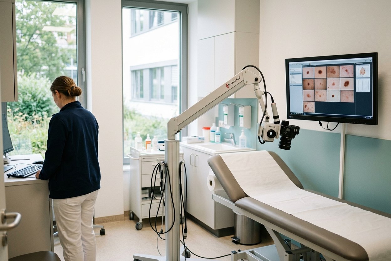
After melanoma, the whole skin and the lymph nodes need regular checks, because both a recurrence and a new melanoma elsewhere are possible. The doctor examines all the skin, not only the scar, feels the nearby nodes and, unless the tumour was very thin and not ulcerated, adds an ultrasound. This is part of the plan, not a formality. A mole changing in shape, border, colour or size over time needs attention; see mole screening and dermatoscopy.
At home, check your skin, scar and nearby nodes regularly, with a mirror or a relative’s help for your back; routine visits can be with a local dermatologist. Worth rechecking in Germany is whatever depends on experience and comparison: lymph node ultrasound, scans in advanced stages, doubtful skin changes. If something changes, do not wait for the next visit. Sun protection is part of the plan, not an afterthought: it lowers the risk of a new skin tumour. The doctor sets the intervals from tumour depth and stage.
Frequently asked questions
Can melanoma be cured for good?
Often, yes. Most melanomas found at stage I or II, before they have spread, are cured by complete removal. At stage III the aim is still cure, with drugs after surgery lowering the risk of recurrence. With distant metastases, immunotherapy and targeted drugs control the disease for a long time in some patients, and some survive it. Individual chances depend on the stage and test results.
What treatment is given for melanoma?
It depends on the stage and the tumour tests. Early melanoma is removed with a margin of healthy skin, often the only treatment needed. A deep tumour or affected nodes call for immunotherapy after surgery, or targeted therapy if the BRAF gene is changed. With distant metastases these drugs are the core, and surgery or radiotherapy treats single deposits. The tumour board sets the plan.
Is a sentinel lymph node biopsy always needed?
No. It is done when the tumour is deep enough or shows risk features such as ulceration, and other tests have found no metastases. A very thin melanoma without such features usually does not need it: node involvement is unlikely, and the operation has its own complications. The decision rests on the histology report, so accurate depth measurement at the slide review matters.
How AlenMed arranges treatment
AlenMed sends your records to the dermato-oncology departments of university hospitals and accompanies you throughout. These include the Clinic for Dermatology, Venereology and Allergology at the University Hospital Frankfurt (headed by Prof. Bastian Schilling), the University Skin Clinic in Tübingen, the Clinic for Dermatology and Venereology at Cologne University Hospital (headed by Prof. Esther von Stebut) and the Clinic for Dermatology at Essen University Hospital. The company has 86 partner hospitals in 38 cities, 345 doctors and its office in Munich.
AlenMed translates reports, sends slides and tissue blocks for review, books appointments, issues the visa invitation, meets you on arrival and provides a medical interpreter. The clinic issues the estimate after reviewing the records; what makes up the bill is explained under melanoma treatment costs.
This material is for information only, is not a public offer and does not replace a consultation with a doctor. Whether treatment is possible, its scope and its outcome are decided only after an in-person examination at the clinic.
News on this topic
Charité treats first beta-thalassemia patient with CRISPR
At Berlin Charité, a 19-year-old beta-thalassemia patient became the first in Germany to receive the newly approved CRISPR gene therapy Exagamglogene Autotemcel.
Real-World Breast Cancer Study Confirms High Five-Year Survival Rates in Germany
A landmark study of over 43,000 German patients reveals a five-year invasive disease-free survival rate of 82.9 percent in early hormone receptor-positive breast cancer.
FDA Approves Jülich Diagnostic Radiotracer FET for Brain Tumor Imaging
The FDA approves the diagnostic radiotracer FET (Pixclara®) for evaluating brain gliomas, highlighting decades of neuro-oncology research at Forschungszentrum Jülich.