EN
Glioma treatment in Germany starts with an exact diagnosis, because glioma is not one disease: the name covers tumours of the brain’s supporting cells, from slow-growing ones people live with for years to aggressive glioblastoma. Almost every decision depends on the report.
This page explains how a German centre reads the report, what molecular findings change, when surgery is offered and when not, what follows, and what a tumour board needs. For the wider picture, see brain tumour treatment in Germany.
What the grade and the molecular report actually mean
The grade describes how aggressively a tumour behaves; the molecular report says which tumour it is. The 2021 WHO classification has four grades, reflecting behaviour, not size. A low grade means slower growth, not harmlessness: diffuse grade 2 gliomas are still malignant.
Four markers change the plan. An IDH mutation separates astrocytomas and oligodendrogliomas from glioblastoma. A 1p/19q co-deletion, loss of parts of chromosomes 1 and 19, plus an IDH mutation defines oligodendroglioma, usually treated with PCV chemotherapy. MGMT promoter methylation silences a DNA-repair gene and, in glioblastoma, predicts benefit from temozolomide. In children and young adults, H3 alterations mark a distinct grade 4 tumour. By current German practice a report lacking these markers is incomplete, so centres usually request the paraffin blocks.
Two people with “the same” diagnosis may therefore be treated differently: location near speech or motor areas, age, general condition and the extent of earlier surgery all matter. Classification and work-up follow the German S2k guideline on gliomas, now under revision.
Diagnosis: imaging, biopsy and neuropathology
Diagnosis rests on imaging, tissue and its analysis. German departments expect MRI without and with contrast to a tumour protocol, if needed with perfusion and MR spectroscopy to locate the most active areas. Scans on an old disc without the required sequences are usually repeated.
If the tumour can be removed safely, tissue comes from the operation; if not, owing to its location or the patient’s condition, a stereotactic biopsy takes small samples through a small hole in the skull. A tumour board of several specialists, not one surgeon, decides, weighing risk against benefit.
The neuropathologist makes the final diagnosis. A frozen section during surgery only confirms that tumour was sampled; paraffin processing takes days, and molecular tests add more, as DNA must be extracted and genes analysed. Planning starts properly only with the full report; if markers are missing, a second opinion from a German doctor can review slides and blocks.
Surgery: how much can be removed safely
Surgeons remove as much tumour as possible without costing speech, movement or vision: surgery alone cannot cure a diffuse glioma, and new deficits can delay further treatment. Yet the extent of removal may affect the outlook and radiotherapy planning, so, as usual in neurosurgery in Germany, functional areas are mapped beforehand and monitored throughout.
Neuronavigation works like GPS on the MRI, showing where the instrument is. Intraoperative monitoring checks with small electrical pulses that motor pathways still work. With 5-ALA, used for malignant gliomas, the patient drinks a solution beforehand and the tumour glows red under blue light. Intraoperative MRI or ultrasound shows whether removable tumour remains. Near the speech area, surgeons may operate with the patient awake, guided by their answers.
These partner departments are examples, not a ranking. Neurosurgery at TUM Klinikum rechts der Isar in Munich is headed by Professor Bernhard Meyer; its focus areas include brain tumour surgery and clinical neuro-oncology. At Helios Klinikum Erfurt, Professor Rüdiger Gerlach heads neurosurgery and the hospital’s neuro-oncology centre; his profile lists surgery in critical areas under neuronavigation and intraoperative monitoring. At Essen University Hospital, Professor Ulrich Sure heads neurosurgery and spinal surgery. For costs, see cost of brain tumour surgery in Germany.
After the operation: radiotherapy, drug treatment and follow-up
Surgery is almost never the whole treatment, because a diffuse glioma grows into the surrounding brain. Grade and molecular findings decide what follows: radiotherapy, chemotherapy, often both, or, for some grade 2 tumours after complete removal, watching with regular scans.
Patients ask most about tumour-treating fields (TTFields), which the German guideline recommends discussing in glioblastoma after chemoradiotherapy. For IDH-mutant grade 2 gliomas not needing radiotherapy or chemotherapy straight after surgery, the EU has approved the targeted drug vorasidenib; most other targeted drugs are still in trials. Immunotherapy remains experimental: in large glioblastoma trials, neither a vaccine nor a checkpoint inhibitor prolonged survival. Always ask: standard care or a study?
Follow-up is part of treatment, not an afterthought: MRI is repeated at set intervals, usually every few months. Early after radiotherapy a lesion can enlarge without tumour growth (pseudoprogression), so a doubtful scan is repeated before treatment changes. Dexamethasone, a steroid for brain swelling, is given only for symptoms and briefly; anti-seizure drugs only after seizures, not for prevention. More on brain tumours and aftercare: Krebsinformationsdienst of the German Cancer Research Center.
Frequently asked questions
How is a glioma different from a glioblastoma?
Glioblastoma is the most aggressive glioma. The WHO reserves the name for diffuse adult tumours without an IDH mutation that show high-grade features; it is always grade 4. IDH-mutant astrocytomas, oligodendrogliomas and pilocytic astrocytomas usually grow more slowly and are treated differently.
Can a glioma be cured?
It depends on the type, and no one can promise an outcome. Circumscribed gliomas such as pilocytic astrocytoma are treated mainly by complete removal, where safe. Diffuse gliomas grow into surrounding tissue and cannot be cured by surgery alone, so radiotherapy, drugs or monitoring follow.
Is radiotherapy always needed after surgery?
No. It is standard in glioblastoma and usually in grade 3 tumours. After complete removal of a grade 2 IDH-mutant glioma, and in younger patients sometimes after incomplete removal, doctors may suggest regular MRI monitoring or vorasidenib tablets instead. The tumour board decides.
How AlenMed organises treatment
With AlenMed, glioma treatment in Germany starts with paperwork: a medical translator prepares reports, neuropathology with blocks or slides and MRI discs for two or three departments. Each reply states whether the patient is accepted, what is proposed and in what order, and includes a cost estimate (see cost of brain tumour treatment in Germany). Papers for a medical visa are prepared in parallel.
AlenMed works with 86 partner hospitals in 38 cities and 345 doctors; the company office is in Munich. A coordinator books appointments, interprets at consultations and on the ward, and collects the discharge letter with the plan for further treatment.
This material is for information only and is not an offer. The treatment plan is decided by a doctor at a face-to-face appointment.
News on this topic
Charité treats first beta-thalassemia patient with CRISPR
At Berlin Charité, a 19-year-old beta-thalassemia patient became the first in Germany to receive the newly approved CRISPR gene therapy Exagamglogene Autotemcel.
Real-World Breast Cancer Study Confirms High Five-Year Survival Rates in Germany
A landmark study of over 43,000 German patients reveals a five-year invasive disease-free survival rate of 82.9 percent in early hormone receptor-positive breast cancer.
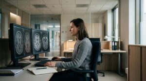
FDA Approves Jülich Diagnostic Radiotracer FET for Brain Tumor Imaging
The FDA approves the diagnostic radiotracer FET (Pixclara®) for evaluating brain gliomas, highlighting decades of neuro-oncology research at Forschungszentrum Jülich.