EN
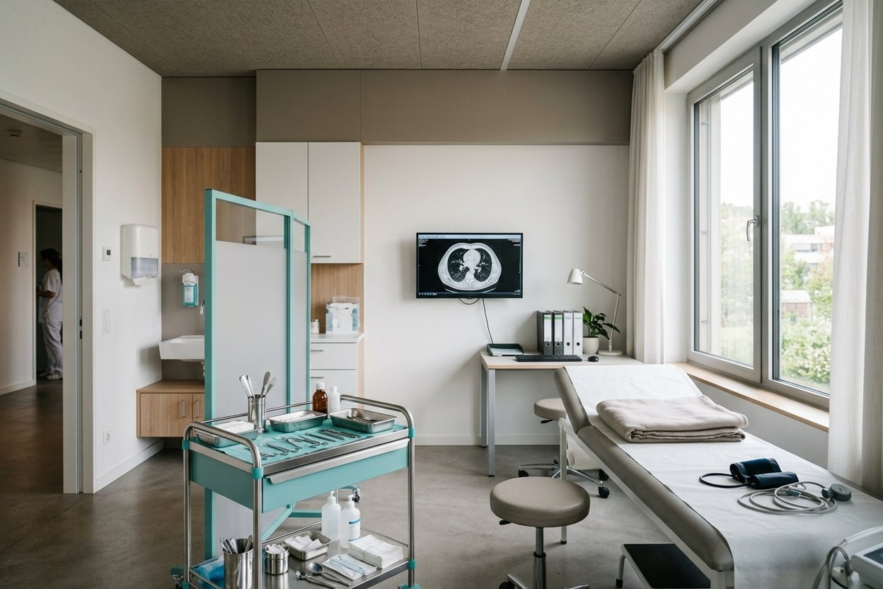
In Germany, lung metastases are treated with surgery, targeted radiotherapy or drugs, often combined, depending on the number of lesions and the original cancer. They usually show up on follow-up CT after bowel, kidney or breast cancer, melanoma or sarcoma. The lungs are a common site because all venous blood passes through their tiny capillaries, where tumour cells get caught.
When there are few lesions, they can be removed or precisely irradiated; treatment of lung metastases in Germany is planned by a tumour board of thoracic surgeons, oncologists, radiologists and pulmonologists. This is not primary lung cancer (see lung cancer treatment): the nodules consist of cells from the original tumour and are treated by its rules. Below: surgery, the tests before it and the alternatives.
When lung metastases are treated locally
Lung metastases are treated locally when there are only a few and the rest of the disease is under control, a situation called oligometastatic disease, from the Greek for “few”. The aim is lasting control, not a promised cure: the disease may stay quiet for a long time, or new nodules may appear.
The board checks that the primary tumour is controlled, that no uncontrolled disease exists in other organs and that the lungs will cope with breathing after surgery. With several organs involved, a staged plan is possible, for example lungs first, then liver (see liver metastases treatment).
Tests before surgery
Doctors first need to know how many lesions there are, where they lie and whether disease is present elsewhere. The basis is a thin-slice chest CT: a chest CT scan with contrast costs approximately 550 euros (an indicative price). PET-CT shows especially active cells, helping to tell a living deposit from a scar or old inflammation and to find disease outside the lungs, though fresh inflammation can light up too.
If the nature of a nodule must be confirmed (metastasis, new lung cancer or benign lump), a tissue sample is taken. In bronchoscopy with endobronchial ultrasound (EBUS), a thin scope with an ultrasound probe is passed into the airways, and a fine needle takes cells from lymph nodes and lesions beside the bronchi. At Asklepios Lungenklinik Gauting, bronchoscopy is led by Dr Wolfgang Gesierich, a pulmonologist and the hospital’s medical director. Molecular tests follow the type of primary tumour and guide the choice of drugs.
Finally, lung reserve is measured: spirometry shows how much air a person breathes out and how fast, diffusion capacity how easily oxygen enters the blood, and scintigraphy how blood flow is spread across the lungs. This shows how much tissue can be removed while leaving enough for breathing.
How the deposits are removed: thoracoscopy, laser, segmentectomy
Deposits are removed whole while sparing as much healthy lung as possible, often by thoracoscopic surgery (VATS) through two or three incisions of one to three centimetres. Compared with open thoracotomy, a cut between the ribs, this means less pain and blood loss and a quicker recovery. It does not suit everyone: large, deep or numerous lesions may need open surgery.
A laser with a wavelength of 1318 nanometres lets the surgeon shell nodules out: the beam cuts while sealing small vessels and airways, so only a thin rim of healthy tissue is lost. This matters when there are many nodules in both lungs.
Near large vessels or bronchi, an anatomical unit is removed instead: a segment (segmentectomy) or a lobe (lobectomy). A frozen section, a quick microscope check of the margins during surgery, confirms that no tumour is left (R0). A systematic review of pulmonary metastasectomy lists complete removal among the main favourable factors, along with control of the primary tumour and few metastases. Surgeons in this field include Stephan Trainer, chief of thoracic surgery at Agaplesion Markus Hospital in Frankfurt, whose department includes lung metastasis surgery, and Dr Rainer Sätzler, chief of thoracic surgery at Klinikum Stuttgart. The operation is described under resection of lung metastases.
When surgery is not possible: radiotherapy and drugs
When surgery is not possible, the main option is stereotactic body radiotherapy (SBRT): a high, tightly focused dose delivered to a small lesion over a few sessions. The machine allows for breathing movement (CyberKnife, for example, tracks the lesion), and no incision or anaesthetic is needed. For some patients the result may be close to surgery: a meta-analysis found no difference in survival between matched patients, although operated patients lived longer overall and randomised trials are lacking.
Drugs follow the primary tumour: chemotherapy, targeted therapy aimed at a specific fault in cancer cells, or immunotherapy, which helps the immune system recognise the tumour. They are often combined with local treatment, which removes visible lesions while drugs act on cells too small to see.
Frequently asked questions
Can lung metastases be cured?
Sometimes, but no doctor can promise it. If there are few lesions, the primary tumour is controlled and all lesions are removed completely, the disease stays away for a long time in some patients. In others new nodules appear, so regular follow-up CT scans are needed, and surgery or radiotherapy may be considered again.
How many lung metastases can be removed?
What matters is not the number but whether all lesions can be removed completely while leaving enough lung for breathing. A laser helps with multiple nodules, even on both sides, as it removes each one with little loss of healthy tissue. The outlook is generally better with few metastases appearing long after the primary tumour was treated.
Surgery or radiotherapy: which is better?
There is no single answer: the tumour board decides based on the number, size and position of the lesions, lung and heart function and the type of primary tumour. Surgery provides tissue for examination and confirms complete removal. Radiotherapy needs no incision and suits patients who cannot have an operation. For some patients, both options are reasonable.
How AlenMed arranges treatment of lung metastases
AlenMed starts with the documents: CT and PET-CT discs, primary tumour treatment records and the histology report. These are translated and sent to the tumour board of partner hospitals; the hospital then issues a cost estimate (Kostenvoranschlag), which depends on the extent of surgery, intensive care days and drugs.
AlenMed also arranges the visa invitation, pick-up and transfer, a medical interpreter at consultations and translation of the discharge report. Its office is in Munich; partner hospitals are across Germany.
This is reference material, not an offer; prices are indicative. Whether surgery is possible is decided by the thoracic surgeon after reviewing the images and seeing the patient in person.
News on this topic
Tübingen researchers uncover mechanism of immune suppression during cancer cell death
Researchers at Universitätsklinikum Tübingen discovered that dying cancer cells release PGE2 to suppress immune response during ferroptosis, opening paths for combination therapy.
New single-cell lung cancer atlas LuCaS+ uses AI to predict metastasis risk
German researchers from LMU, Charité, and TU Berlin created the LuCaS+ single-cell atlas, using AI to predict lung cancer metastasis risk from archived tissue samples.
ForTra Funds Clinical Trials for Leukemia and Muscular Dystrophy
ForTra gGmbH provides four million euros to support clinical trials for acute myeloid leukemia and Duchenne muscular dystrophy in Germany.