EN
Brain cancer treatment in Germany is provided by neuro-oncology centres at large hospitals: they first establish what the tumour is and exactly where it lies, then a tumour board chooses surgery, radiotherapy, drugs or a combination. Most people arrive with an MRI scan, an alarming report and pressure to decide fast — yet the brain carries speech, movement and memory, so haste is the main risk.
A German work-up keeps a strict order: first the nature and exact location of the tumour, then the choice of method. That promises no particular outcome, but it fits the treatment to this tumour rather than to the label “brain tumour”.
Which brain tumours are treated in Germany
German hospitals treat tumours of the brain and its coverings — gliomas, including glioblastoma, and meningiomas — as well as metastases from cancers elsewhere. Brain tumour treatment in Germany starts with naming the tumour: benign or malignant? A benign tumour usually grows slowly without invading nearby tissue and, if well placed, can often be removed completely. A malignant one grows faster and invades the brain around it, so surgery alone is rarely enough; everything else follows from this.
Among tumours of the brain tissue itself, gliomas are the most common: they grow from glial cells, the brain’s supporting tissue, and vary in how malignant they are (see glioma treatment). Glioblastoma, the most aggressive glioma, grows fast and spreads into neighbouring tissue (see glioblastoma treatment). Meningioma grows from the membranes covering the brain and is usually benign.
Brain metastases are a different task from a primary tumour. “Primary” means the tumour started in the brain or its coverings; metastases are cells from a cancer in another organ, most often the lung, breast or kidney, or a melanoma. So the plan is made with the oncologist treating the original cancer.
Getting the diagnosis right
The diagnosis comes in two steps: imaging shows where the tumour is, tissue analysis what it is. The main method is MRI, including special sequences that show not only the shape of the tumour but how it relates to the speech and motor areas and the nerve pathways. Where needed, PET (positron emission tomography) adds a measure of tissue activity, helping to tell the most aggressive parts of the tumour from quieter ones.
But a scan shows where the tumour is and how it behaves, not what it is at cell level. Only tissue gives that answer, so a biopsy is often taken — sometimes stereotactically, with equipment calculating the path to a deep lesion and a sample taken through a small opening. A pathologist examines the tissue, and molecular testing is now always added: brain tumours have genetic features that determine both the exact diagnosis and the drug options.
Before a consultation, have the MRI on disc rather than as printouts, which lose the special sequences and fine detail. After surgery or a biopsy, the doctor also needs the slides and paraffin blocks with the pathology report, so the diagnosis can be checked and missing molecular tests done.
Surgery and what makes it precise
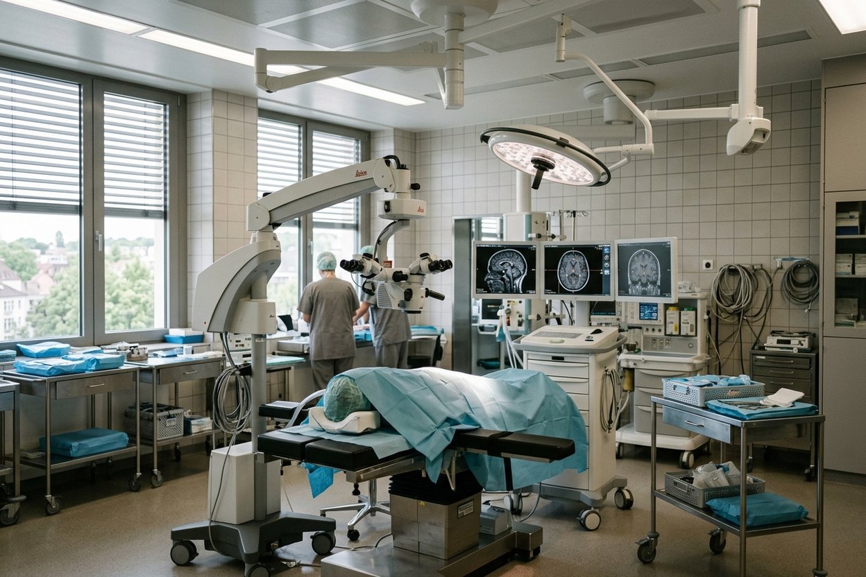
Precision comes from brain mapping, function monitoring and techniques that show the tumour border. If the tumour can be reached safely, surgery usually comes first, aiming to remove as much tumour as possible without harming speech and movement. Neuronavigation guides the surgeon along a map built in advance, and function is monitored throughout — sometimes the patient is woken for part of the operation to confirm that speech and movement are intact. Intraoperative MRI, scanning during the operation, and fluorescence guidance show the border: after a dose of 5-ALA, glioma tissue in particular glows under special light. Some tumours near the pituitary gland or in the fluid-filled ventricles are removed with an endoscope via the nose or a small opening in the skull.
Such operations are done by neuro-oncology departments of large hospitals. At Helios Klinikum Erfurt, Prof. Rüdiger Gerlach, chief of neurosurgery, heads the neuro-oncology centre; in Munich, Prof. Bernhard Meyer directs the department of neurosurgery at Klinikum rechts der Isar; in Tübingen, Prof. Marcos Tatagiba heads the university neurosurgery clinic. The LMU Comprehensive Cancer Center in Munich also has a neuro-oncology centre. See also neurosurgery in Germany.
Hospital stay and recovery depend on the extent of surgery and the patient’s condition. A follow-up MRI soon after surgery shows how much tumour was removed. Early rehabilitation starts in hospital with physiotherapy and, if needed, speech therapy; if deficits persist, neurological rehabilitation may follow. MRI checks then continue on a schedule.
Radiotherapy and drug treatment
Radiotherapy and drugs come in when surgery is impossible or not enough. Stereotactic radiotherapy, Gamma Knife and CyberKnife are not separate miracle cures but different ways of delivering a precise dose while sparing surrounding tissue; a small, well-defined lesion such as a metastasis may be treated in one high-dose session, called radiosurgery. Proton therapy is an option for selected cases, above all children and tumours next to critical structures such as the optic nerve; it is given on indication, not as a “more modern” replacement for conventional radiotherapy.
Drug choice follows the molecular profile: chemotherapy or, with a suitable genetic target, targeted drugs. Immunotherapy in Germany for a brain tumour is used only in a narrow set of situations, mostly in clinical studies: it has not beaten standard treatment in glioblastoma, though it may help with brain metastases if it works against the original cancer. Dendritic cell vaccines, made from the patient’s own immune cells, and other experimental offers from private clinics are not standard glioblastoma treatment either. German experts advise their use only within studies; outside one, let the tumour board weigh it as an individual decision.
The neuro-oncology board — a neurosurgeon, an oncologist, a radiation oncologist, a neurologist and a pathologist — decides which methods to combine and in what order, following approved protocols rather than one doctor’s preference. The Krebsinformationsdienst of the German Cancer Research Center offers a clear overview of brain tumours in German.
A second opinion, and the money question
A second opinion is most useful before surgery, while the plan can still change. A German specialist re-reads the scans and any pathology and says whether they agree with the diagnosis and approach, and whether the tumour can be reached with less risk; sometimes that changes the plan. See second opinion from a German doctor.
The clinic sets out the cost in a quote after reviewing the documents, so there are no figures here. It depends on what lies ahead — a scan review, surgery, radiotherapy, drugs or a combination — and on the length of stay; see cost of brain tumour treatment.
Frequently asked questions
Which hospital in Germany is best for brain tumour treatment?
There is no single winner; choose the department that suits your tumour. German experts advise a centre with extensive experience of brain tumours. Signs of one: a large brain tumour caseload, a regular neuro-oncology board, intraoperative MRI or fluorescence guidance, and German Cancer Society certification as a neuro-oncology centre.
Can a brain tumour be cured?
It depends mainly on the tumour type and location. A benign tumour such as a meningioma can often be removed completely if it is easy to reach. Malignant gliomas grow into the surrounding tissue and usually cannot be removed entirely, so treatment aims to hold them back and preserve quality of life.
Is surgery always needed for a brain tumour?
No. If the tumour lies deep or where surgery is too risky, or the patient is frail, doctors often take only a stereotactic biopsy, then use radiotherapy and drugs. Small metastases and some well-defined benign tumours are treated with radiosurgery, without an incision. The tumour board decides.
Can treatment continue in Germany after surgery at home?
Yes, this is common. German doctors review the MRI scans from before and after surgery and the tumour slides, adding missing molecular tests on the paraffin blocks. The tumour board then decides whether more surgery, radiotherapy or drugs are needed, so bring the slides and blocks, not just the discharge report.
How AlenMed organises the treatment
AlenMed matches the clinic and doctor to your documents, has the reports translated and sent to the right department, books appointments, prepares the visa invitation, arranges an interpreter at consultations and stays with the patient throughout treatment.
Its office is in Munich and its partner clinics are all over Germany; the clinic is chosen for the tumour, not for nearness to the office.
This material is for information only and does not replace a consultation with a doctor. Whether treatment is possible, what it will involve and what it will achieve can be determined only after an examination at the clinic in person. The information on this page is not an offer.
News on this topic
Charité treats first beta-thalassemia patient with CRISPR
At Berlin Charité, a 19-year-old beta-thalassemia patient became the first in Germany to receive the newly approved CRISPR gene therapy Exagamglogene Autotemcel.
Real-World Breast Cancer Study Confirms High Five-Year Survival Rates in Germany
A landmark study of over 43,000 German patients reveals a five-year invasive disease-free survival rate of 82.9 percent in early hormone receptor-positive breast cancer.
FDA Approves Jülich Diagnostic Radiotracer FET for Brain Tumor Imaging
The FDA approves the diagnostic radiotracer FET (Pixclara®) for evaluating brain gliomas, highlighting decades of neuro-oncology research at Forschungszentrum Jülich.